Over the years, increasing efforts have been made to reduce the radiation exposure of therapists and patients.
Nowadays, most people and institution relies on trusted Lead Shielding company to design and build radiation reduction equipment.
However, can you imagine how people protect themselves before these companies exist?
Radiation protective clothing
First lead aprons and lead gloves to protect against X-rays, around 1920
After Rollins' discovery in 1920 that lead aprons to protect against X-rays, lead aprons with a lead thickness of 0.5 mm were introduced.
Due to their high weight, lead-free or lead-reduced aprons were subsequently developed. In 2005, it was recognized that the protection was sometimes considerably less than when wearing lead aprons.
The lead-free aprons contain tin, antimony, and barium, which have the property of developing intense natural radiation ( X-ray fluorescence radiation) when exposed to radiation.
The manufacturer must specify the weight per unit area in kg / m² at which the protective effect of a pure lead apron of 0.25 or 0.35 mm Pb is achieved. The protective effect must be suitable for the energy range used, for low-energy aprons up to 110 kV, and for high-energy aprons up to 150 kV.
Lead Glasses
People also needed to use Lead glasses with front glasses with a lead equivalent of 0.5–1.0 mm lead, depending on the application, with a lead equivalent for the side protection of the 0.5–0.75 mm lead.
Outside of the useful beam, the radiation exposure mainly arises from the illuminated tissue's scattered radiation. When examining the head and torso, this scattered radiation can spread inside the body and can hardly be shielded by radiation protective clothing. However, the fear that a lead apron will prevent radiation from leaving the body is unfounded, as lead absorbs radiation strongly and hardly scatters it.
To prepare an orthopantomogram (OPG) for a dental overview, it is sometimes recommended to dispense with a lead apron. This barely shields the scattered radiation from the jaw area but can hinder the rotation of the recording device. [50] According to the X-ray Ordinance valid in 2018, however, it is still mandatory to wear a lead apron when making an OPG.
Calcium tungstate was replaced in the 1970s with luminescent screens (terbium-activated lanthane oxybromide, gadolinium oxysulphide) built from rare earth, much better for amplification and accuracy. The lack of picture clarity has not attracted the use of screen intensification in dental film development. The combination with highly sensitive films reduced the radiation exposure even further.
Anti-Scatter grid
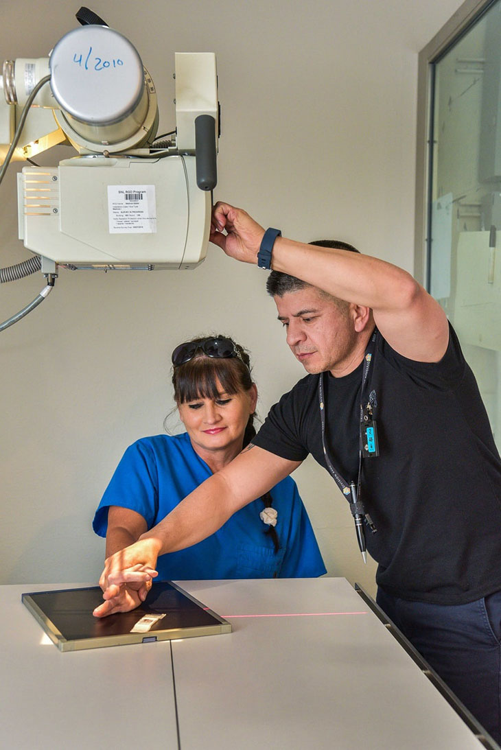
An anti-scatter grid (Potter-Bucky grid) is a device in X-ray technology attached in front of the image receiver (screen, detector, or film). It reduces the incidence of diffuse radiation on it.
The first anti-scatter grid was developed in 1913 by Gustav Peter Bucky (1880–1963). The American radiologist Hollis Elmer Potter (1880–1964) improved it and added a movement device in 1917.
When using anti-scatter grids, the radiation dose must be increased. In children, therefore, the use of anti-scatter grids should be avoided if possible. With digital radiography, a grid can be dispensed under certain conditions to reduce the patient's exposure to radiation.


